





How a Lateral-Flow Test Works
Rapid immunochromatographic tests (also known as lateral-flow tests) were originally developed in the late 1980s for the rapid and straightforward detection of pregnancy. Since then, this method—which requires neither trained personnel nor specialized laboratory equipment, and which is also quick to perform and cost-effective—has become firmly established in point-of-care diagnostics, particularly in human medicine. On-site diagnostics using rapid immunochromatographic test systems are already in use in the veterinary field as well, and we at Fassisi® are continuing to expand and further establish these systems.
Our rapid immunochromatographic test systems typically consist of a sample pad, a conjugate pad, a reaction matrix, and a wicking or absorbent pad.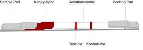
As shown in the figure, the individual components overlap one another and are applied to a special card for fixation.
The sample is first placed on the sample pad, which prepares it for the rest of the test: For example, materials are used here that separate whole blood so that only serum reaches the second component of the test, the conjugate pad. A particulate conjugate has been immobilized on this pad. The particles are mostly gold particles to which a specific biological component (an antigen or an antibody) of the test has been bound.
As soon as the sample comes into contact with the conjugate pad, it dissolves the conjugate, thereby enabling a reaction between the analyte and the first biological component. This complex of the analyte and the first biological component then migrates to the third component of the lateral-flow test: the reaction matrix.
This consists of a porous membrane onto which additional biological components have been applied in the form of a test line and a control line. The test line consists of proteins (antigens or antibodies) that bind the complex of the analyte and the first biological component to the membrane, thereby producing a color signal. Conjugated particles that have not bound to the analyte continue to migrate toward the control line, which typically consists of a species-specific anti-immunoglobulin antibody that reacts with the antibody in the conjugate and always produces a color signal, regardless of the result of the test line. The fourth component of the lateral-flow test is the wicking pad, which absorbs the fluid from the reaction matrix and thus ensures the flow from the sample pad through the conjugate pad and the membrane.
These direct rapid test systems are used when larger analytes with multiple antigenic determinants need to be detected, such as in the detection of parvovirus with our Fassisi® ParCo. In this case, a positive result is indicated by the appearance of both a test line and a control line.
The video demonstrates how a sandwich test works, in which the analyte is present in the sample, causing a color signal to appear on both the test line and the control line.